Facility
Multiscale Imaging

The Multiscale Imaging Unit provides access to state-of-art light microscopy for scientists within and outside the institute, and performs independent and collaborative research for the development of advanced imaging-based technology.
The Multiscale Imaging Unit comprises a wide range of microscopes for live or fixed samples, including in vivo optical imaging instruments for luminescence and fluorescence tomography. High-capacity network of digital workstations is also available for the processing and analysis of imaging data.
The staff is involved in a wide range of scientific collaborations to support users in their studies in different fields of research, such as Oncology, Immunology, Neuroscience and Cardiology. The staff is also focused to the development of new methodologies and techniques aimed at significantly enhance bioimaging research.
major objectives of the Unit
🢒 Set up, maintain and develop optical technology for basic and advanced research
🢒 Provide theoretical and practical training to introduce researchers to optical and advanced imaging applications
🢒 Perform independent and collaborative research for the development of advanced imaging-based technology.
among our technologies
Bright-field microscopes
Wide-field fluorescence microscopes
Confocal microscopes
Super-Resolution (STED) mode
Single-Molecule Detection (SMD, FLIM/FCS) mode
Multi-photon microscope
Luminescence tomography
Fluorescence tomography
Metal-tagged Imaging Mass Cytometry
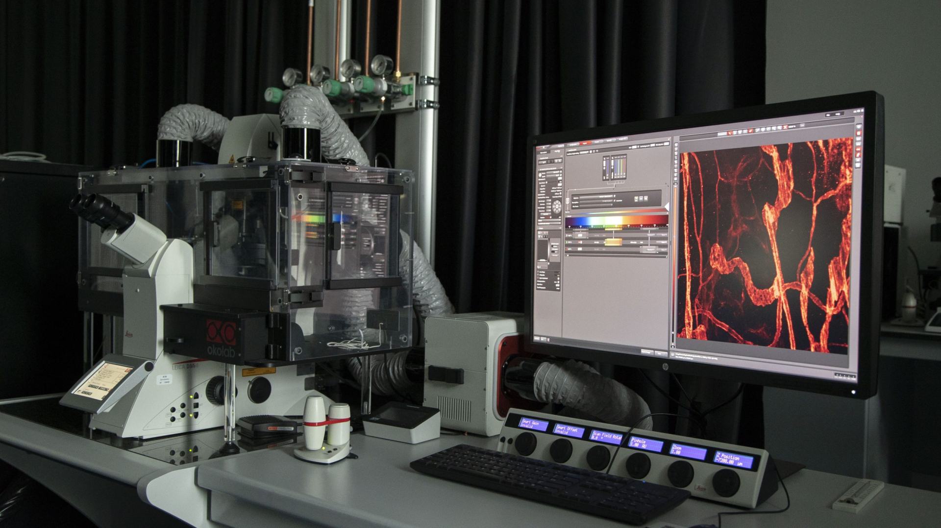
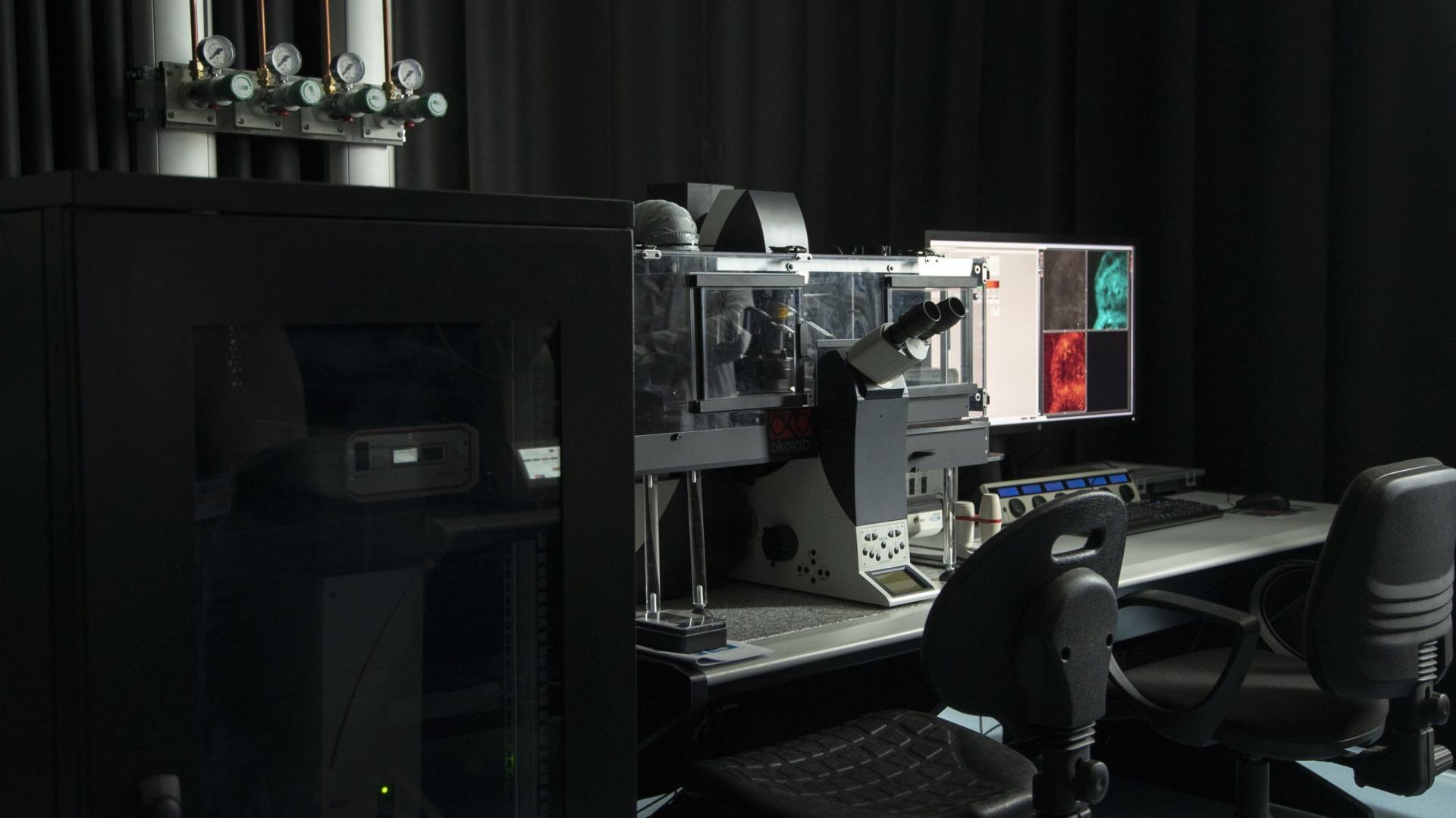
Machinery
We use high end technology of the latest industry standarts
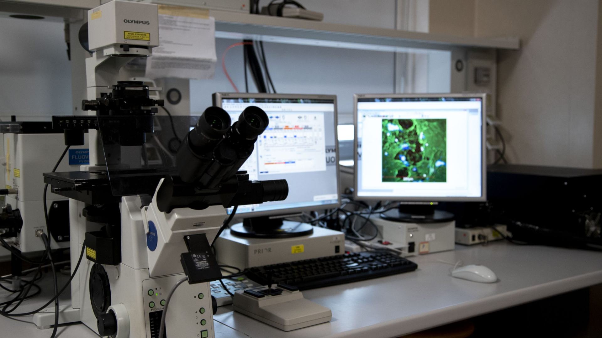
Confocal Microscope FV1000
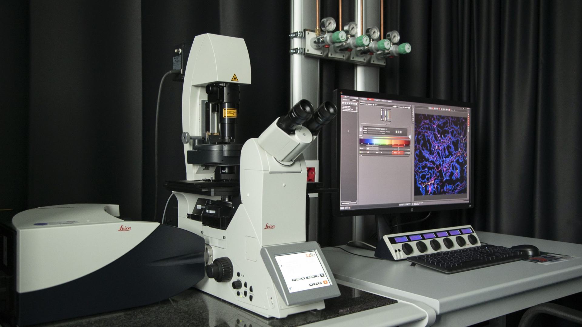
LEICA SP8 Confocal Microscope (Basic)
LEICA SP8 Confocal Microscope (Advanced)
LEICA SP5 Confocal Microscope
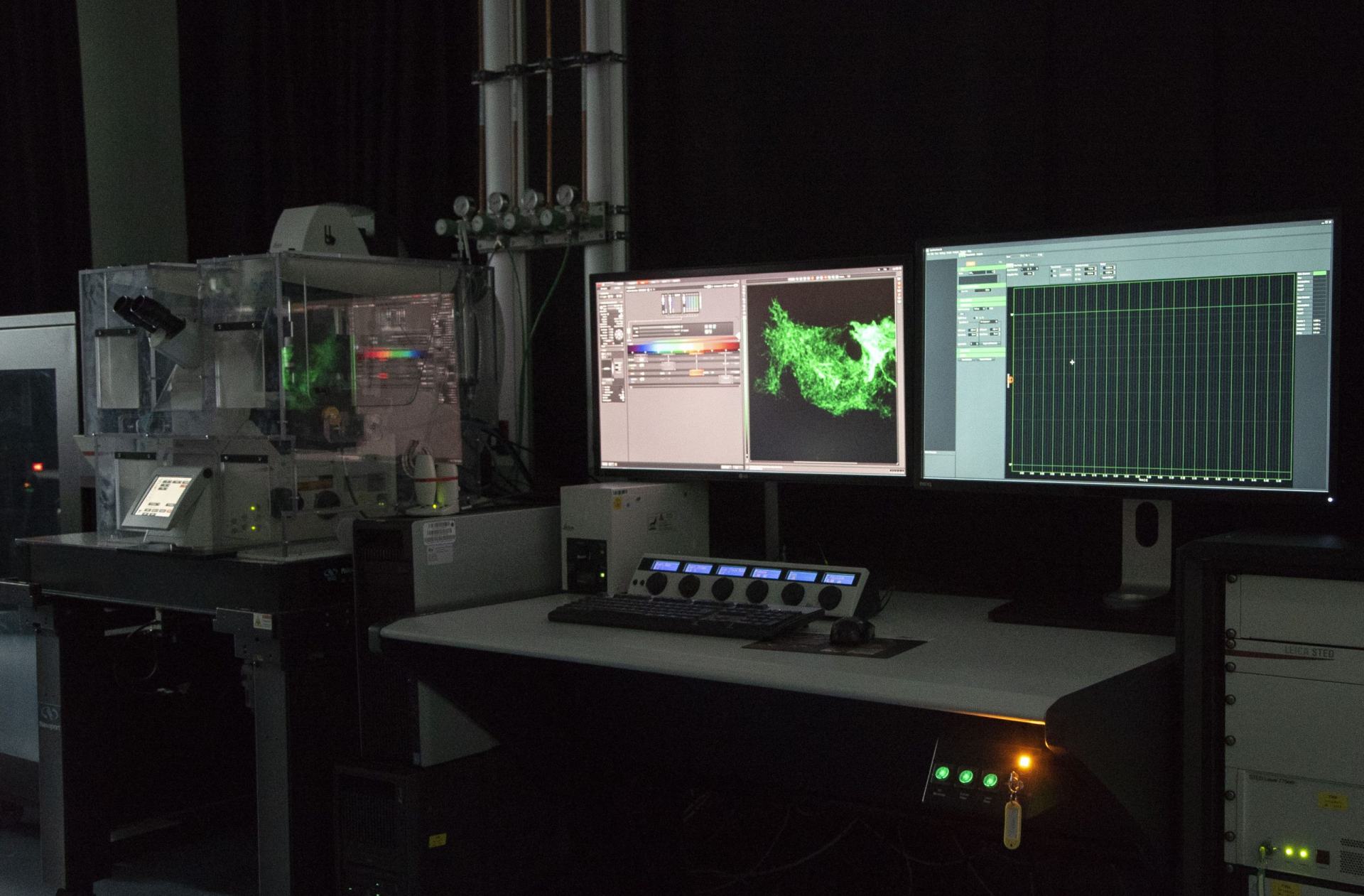
LEICA SP8 STED3X SMD/FCS Confocal Microscope
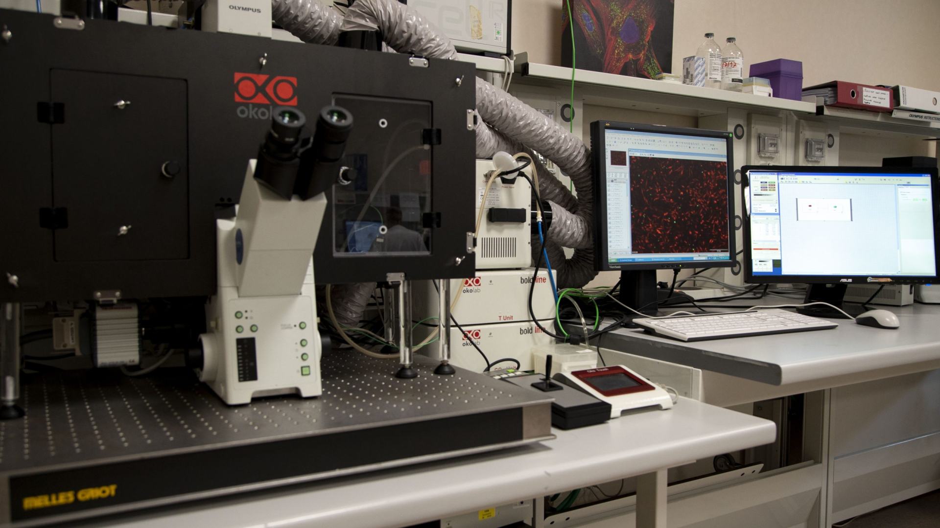
Widefield CellR live cell imaging
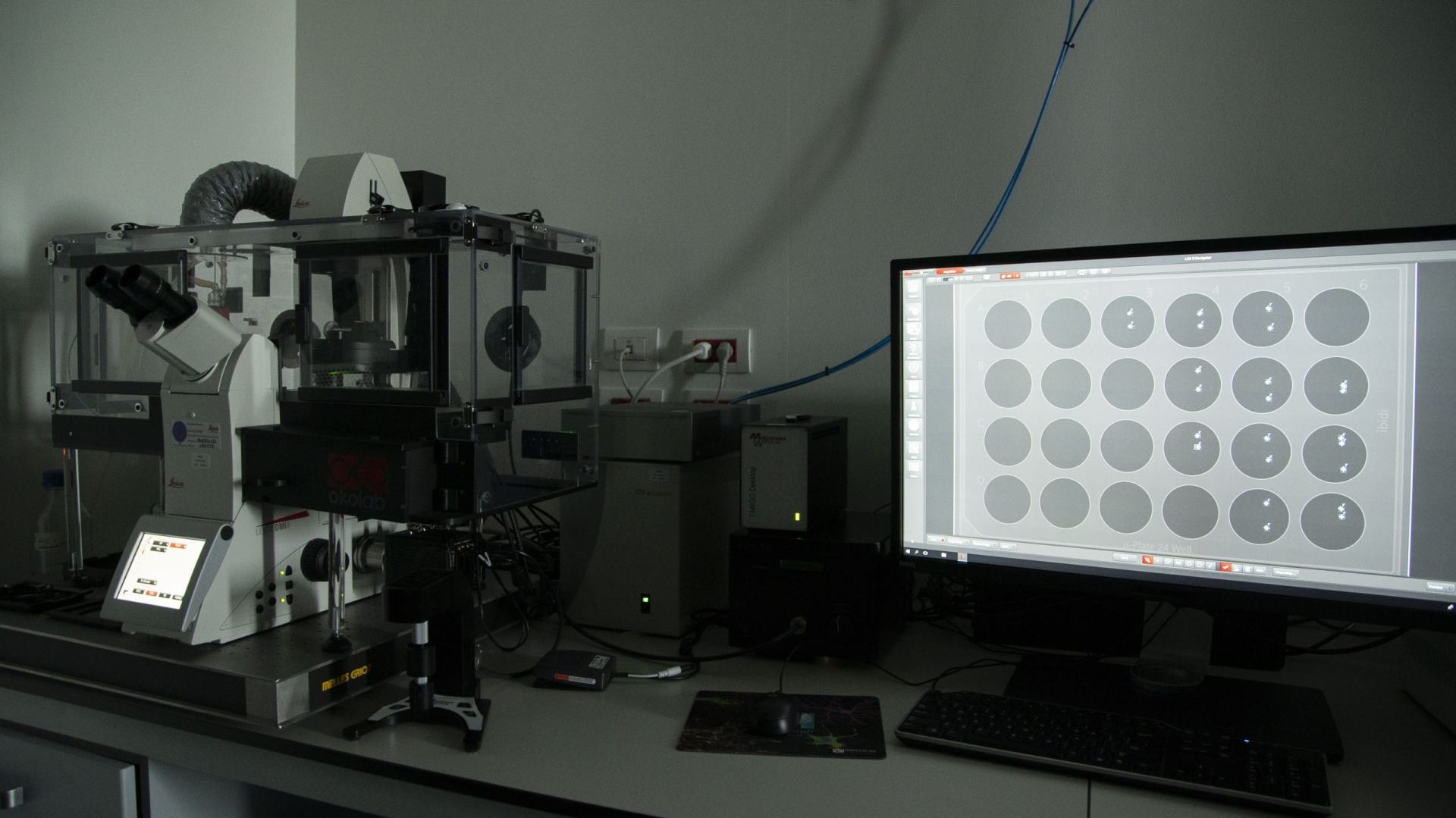
LEICA DMi8 Wide-Field Microscope (Live Cell imaging)
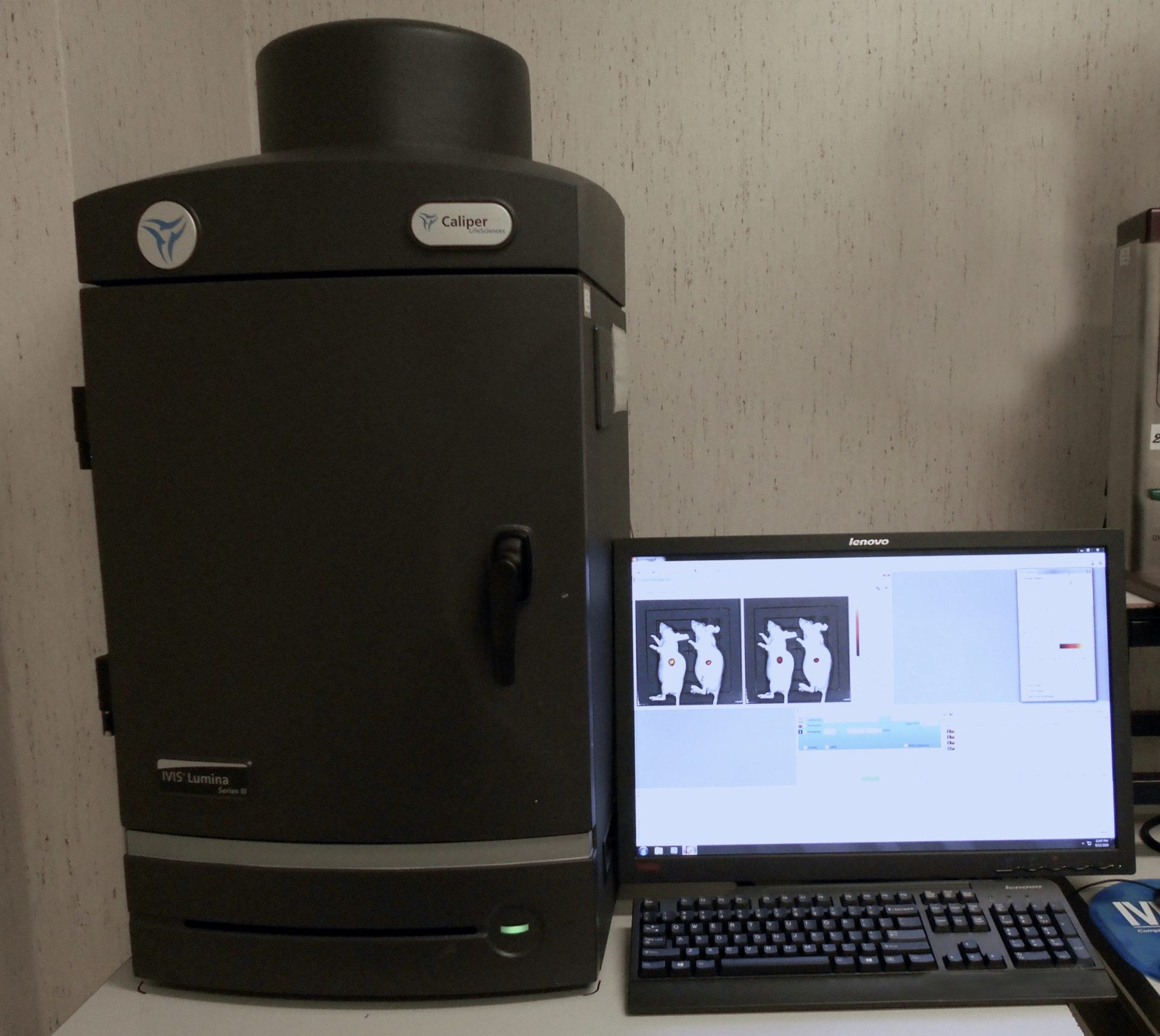
IVIS Lumina III
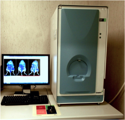
FMT2000 (Fluorescence Tomography system)
Tissue optical clearing system
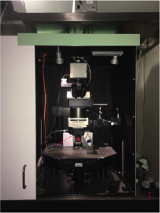
Multiphoton Lavision Trimscope FLIM
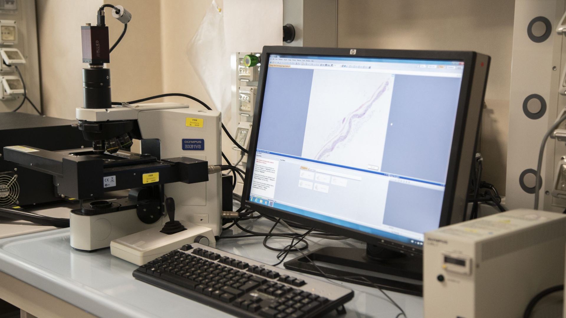
Slide Scanner vs120 dotSlide
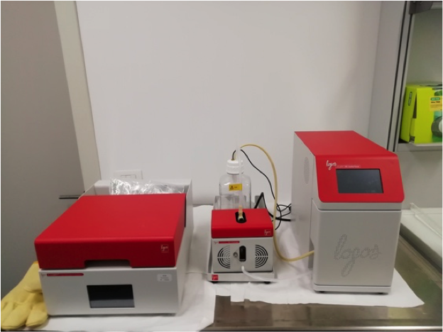
Hyperion™ Imaging System
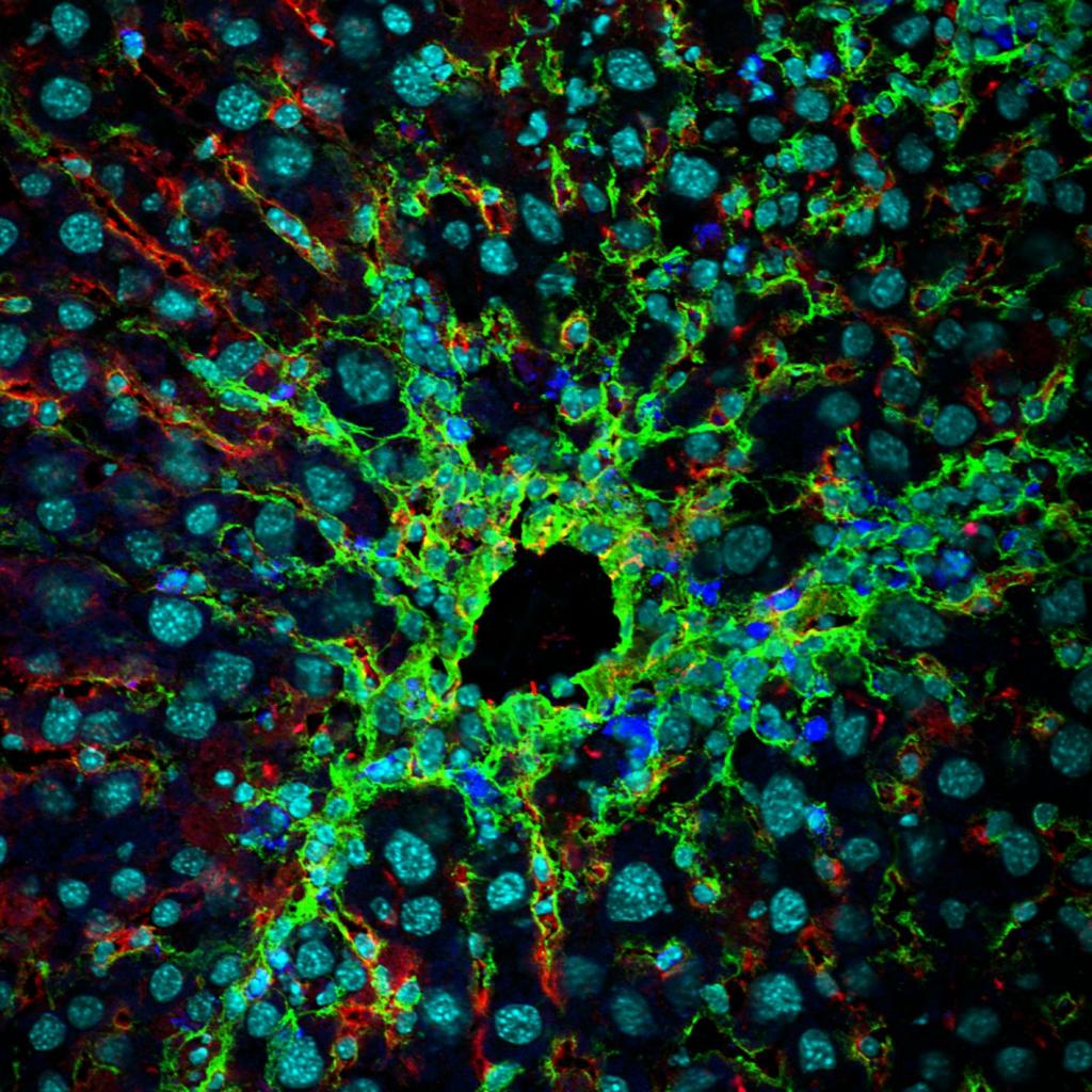
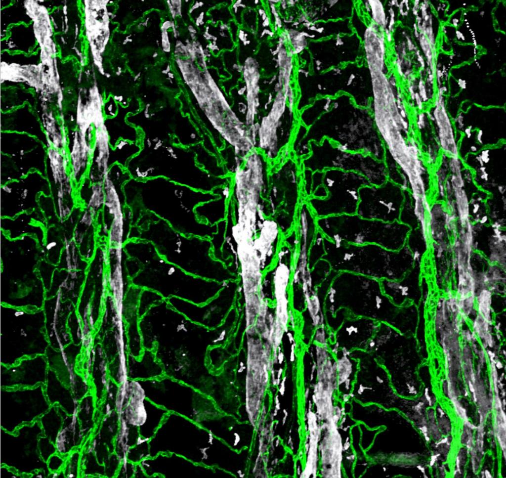
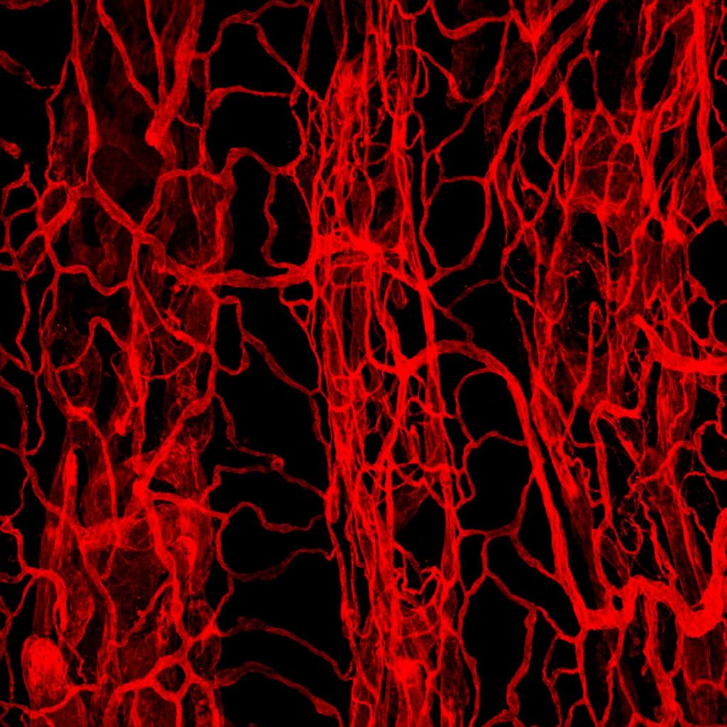
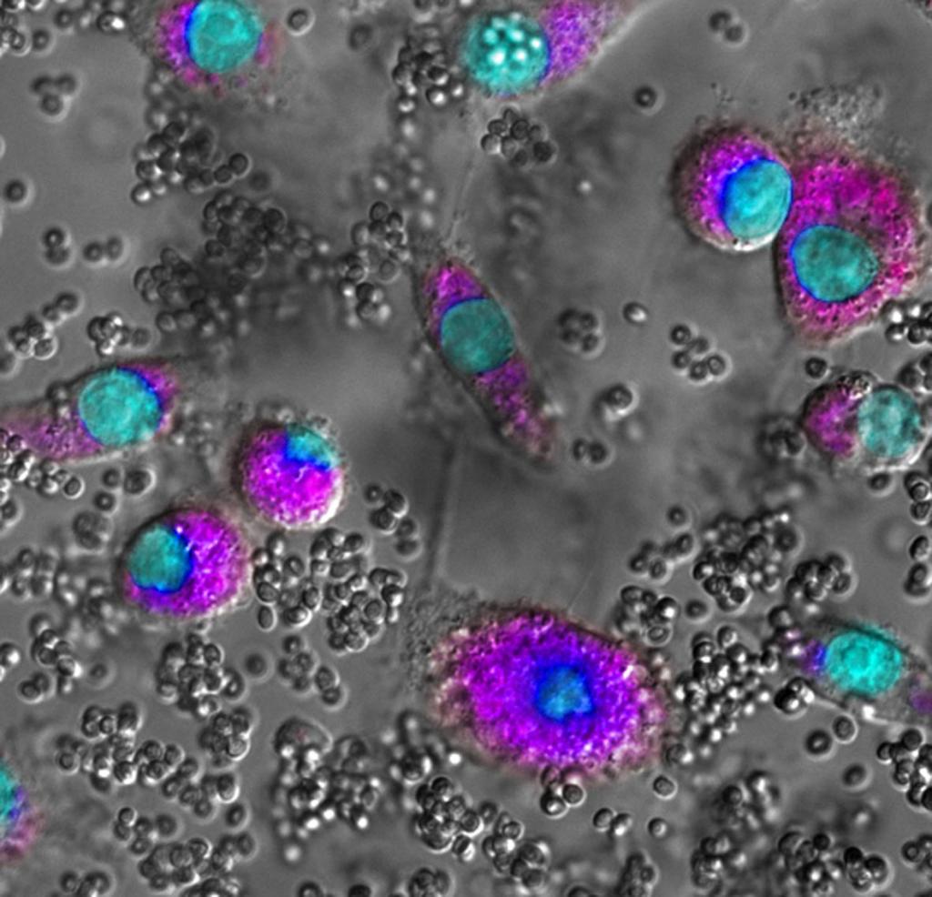
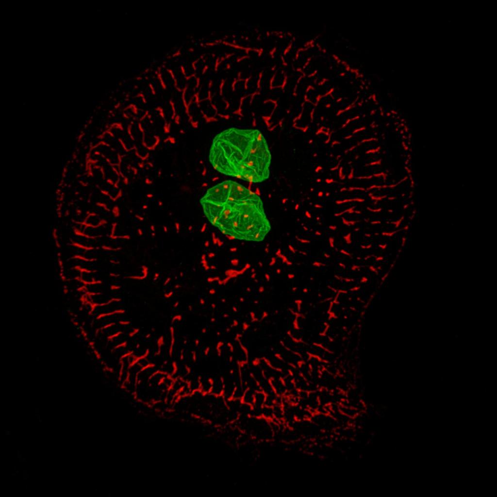
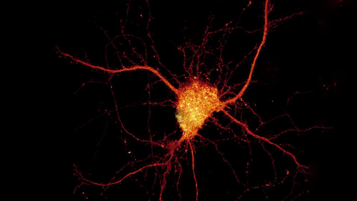
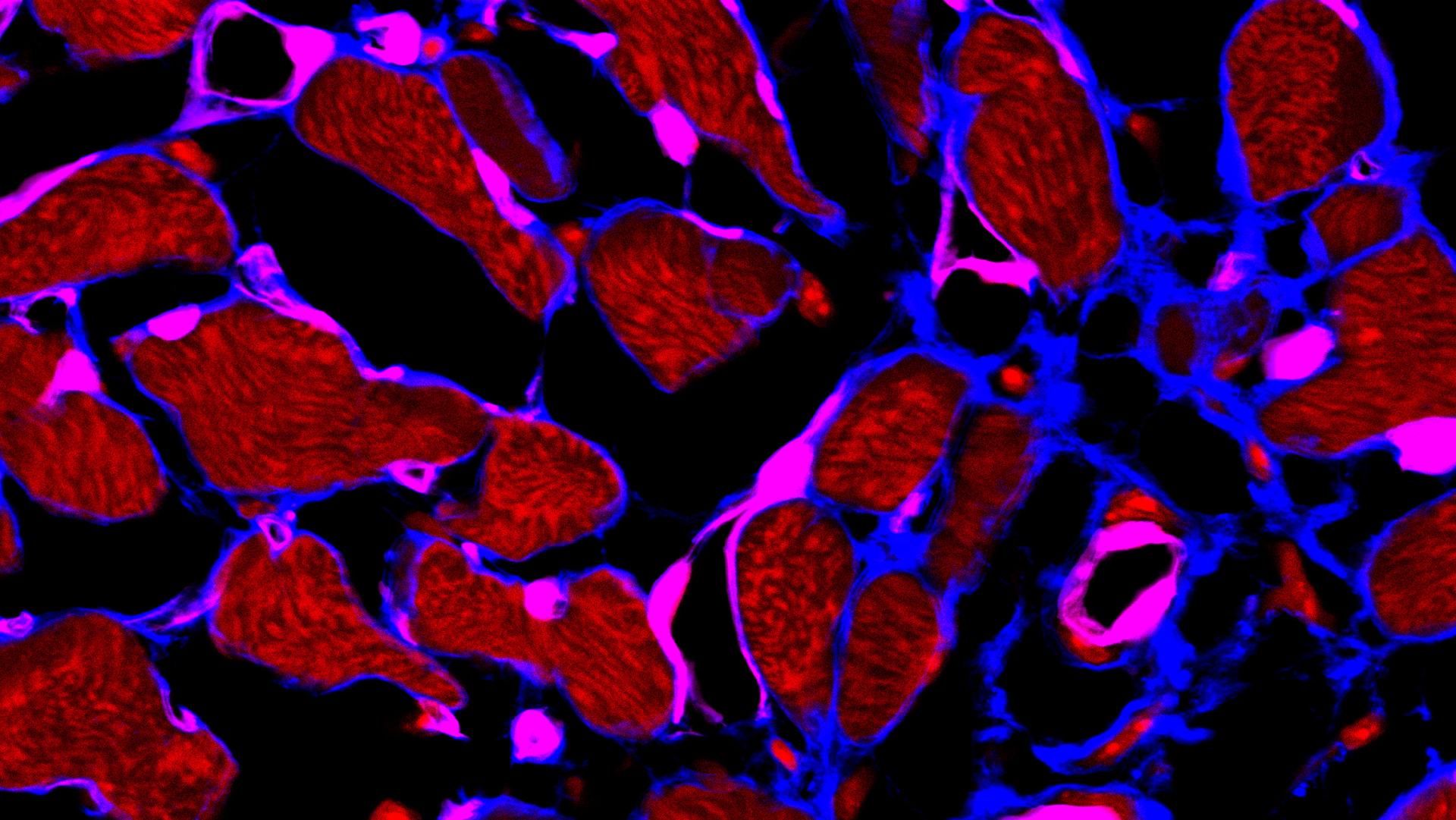
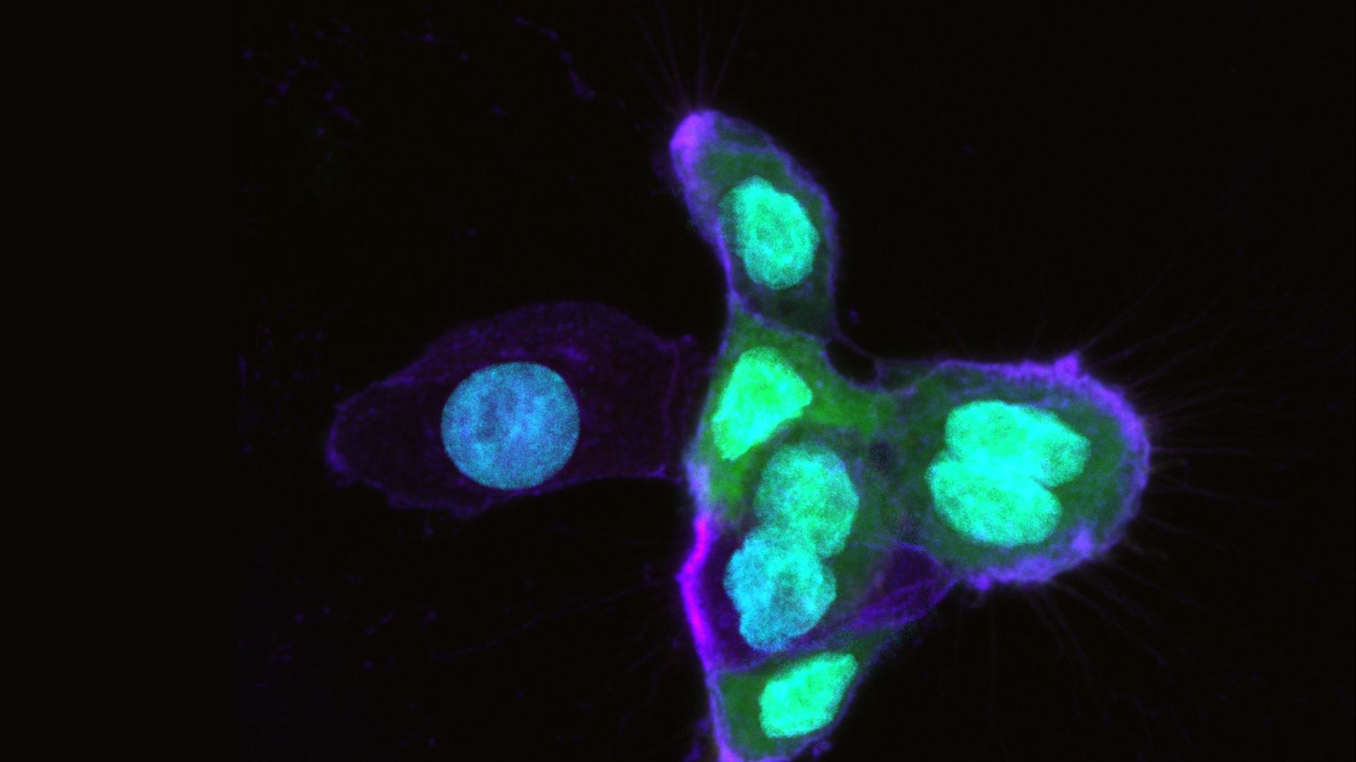
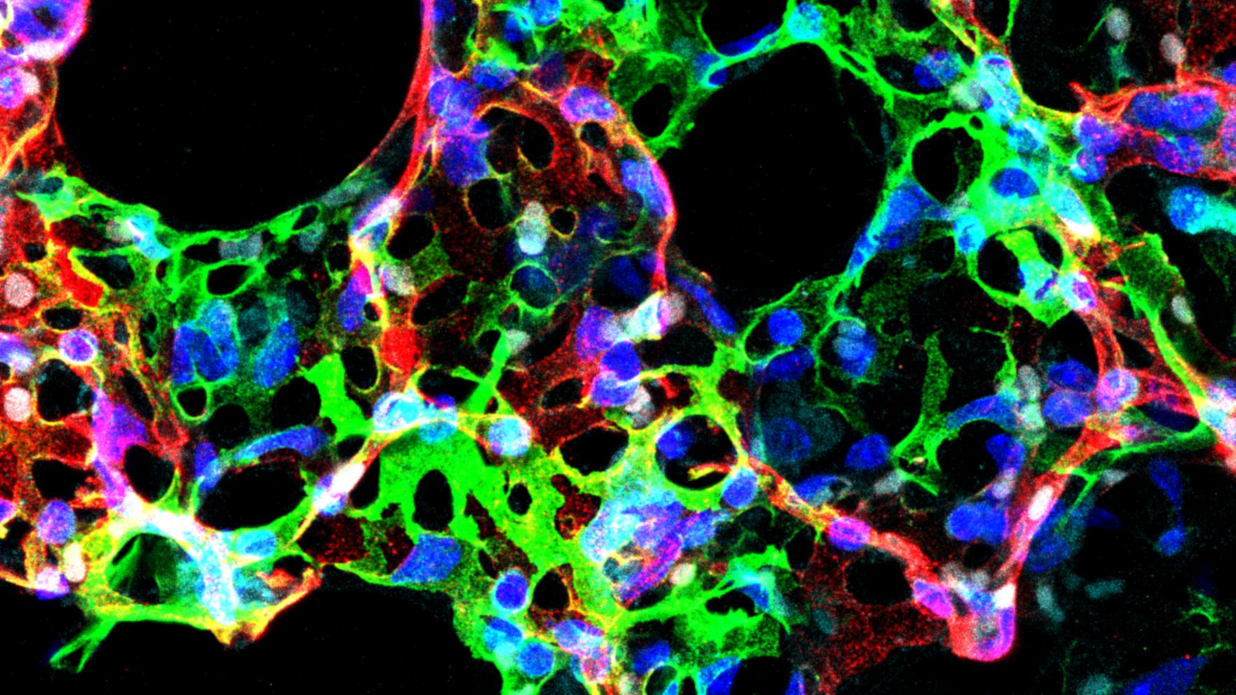
Facility Gallery
Selected publications
Maternal immune activation leads to defective brain-blood vessels and intracerebral hemorrhages in male offspring.
Serum amyloid P component is an essential element of resistance against Aspergillus fumigatus.
Nanoscale Study of Calcium Handling Remodeling in Right Ventricular Cardiomyocytes Following Pulmonary Hypertension.
Nanobodies as Versatile Tool for Multiscale Imaging Modalities.
Local externalization of phosphatidylserine mediates developmental synaptic pruning by microglia.
Optimization of a Luciferase-Expressing Non-Invasive Intrapleural Model of Malignant Mesothelioma in Immunocompetent Mice.
Tumor-Derived Prostaglandin E2 Promotes p50 NF-κB-Dependent Differentiation of Monocytic MDSCs.
Single-Cell Sequencing of Mouse Heart Immune Infiltrate in Pressure Overload-Driven Heart Failure Reveals Extent of Immune Activation.
The macrophage tetraspan MS4A4A enhances dectin-1-dependent NK cell-mediated resistance to metastasis.
Circ_Lrp6, a Circular RNA Enriched in Vascular Smooth Muscle Cells, Acts as a Sponge Regulating miRNA-145 Function.
3D Bone Biomimetic Scaffolds for Basic and Translational Studies with Mesenchymal Stem Cells.
The Microglial Innate Immune Receptor TREM2 Is Required for Synapse Elimination and Normal Brain Connectivity.
Inhalation of peptide-loaded nanoparticles improves heart failure.
IL-1R8 is a checkpoint in NK cells regulating anti-tumour and anti-viral activity.
Identification of human somatostatin receptor 2 domains involved in internalization and signaling in QGP-1 pancreatic neuroendocrine tumor cell line.
Adeno-associated virus-mediated CASQ2 delivery rescues phenotypic alterations in a patient-specific model of recessive catecholaminergic polymorphic ventricular tachycardia.
RORC1 Regulates Tumor-Promoting “Emergency” Granulo-Monocytopoiesis.
An acidic microenvironment sets the humoral pattern recognition molecule PTX3 in a tissue repair mode.
Occurrence of tertiary lymphoid tissue is associated with T-cell infiltration and predicts better prognosis in early-stage colorectal cancers.
The humoral pattern recognition receptor PTX3 is stored in neutrophil granules and localizes in extracellular traps.
Facility Staff